SINGLE ANTERIOR · CASE 08

One Lateral. Every Detail.

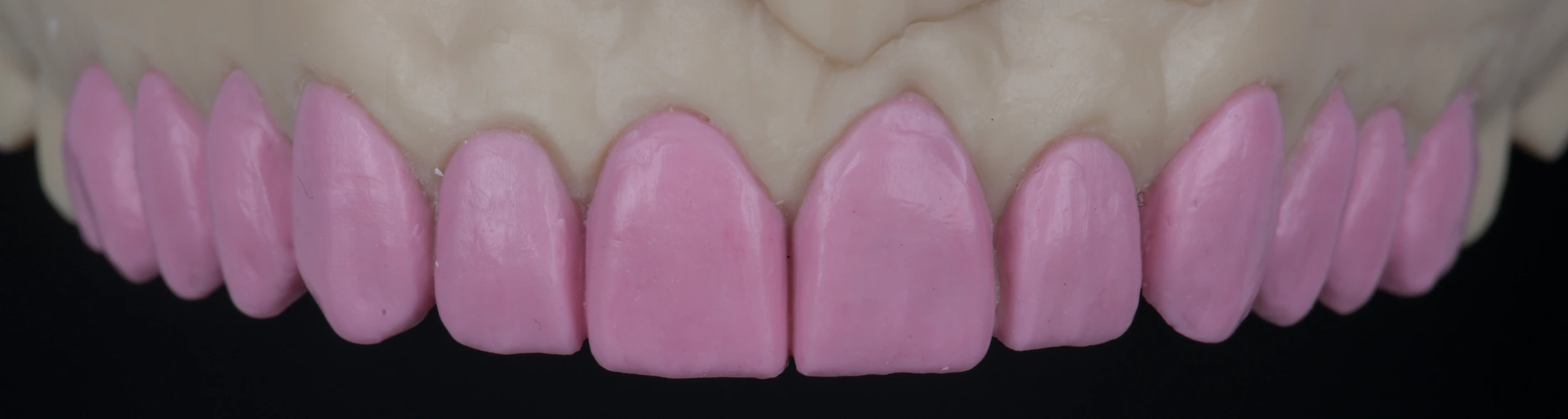
This single lateral-incisor case is documented through baseline smile and anterior views, single-tooth contour planning on a model, and clinical shade-reference photographs. Later smile, frontal and palatal views show the lateral-incisor contour in relation to the surrounding teeth.

Original case photography. Clinical treatment is provided by the treating dentist; individual results vary.

SEE THE DETAILS SIDE BY SIDE

Before & after.

Select any photograph to see the original framing at full size.

Opposite-side retracted lateral view with teeth apart

Before
After

Both views show separated teeth from the same lateral perspective. Photographic lighting, framing and mouth opening vary.

THE PHOTO SERIES

A closer look
at the craft.

01

Starting clinical views

Before: opposite-side retracted lateral view with teeth separated
02

Laboratory planning & interim detail

A pink single-tooth contour pattern on an upper anterior model.
BL4 and B1 shade-reference tabs held beside the upper anterior teeth.
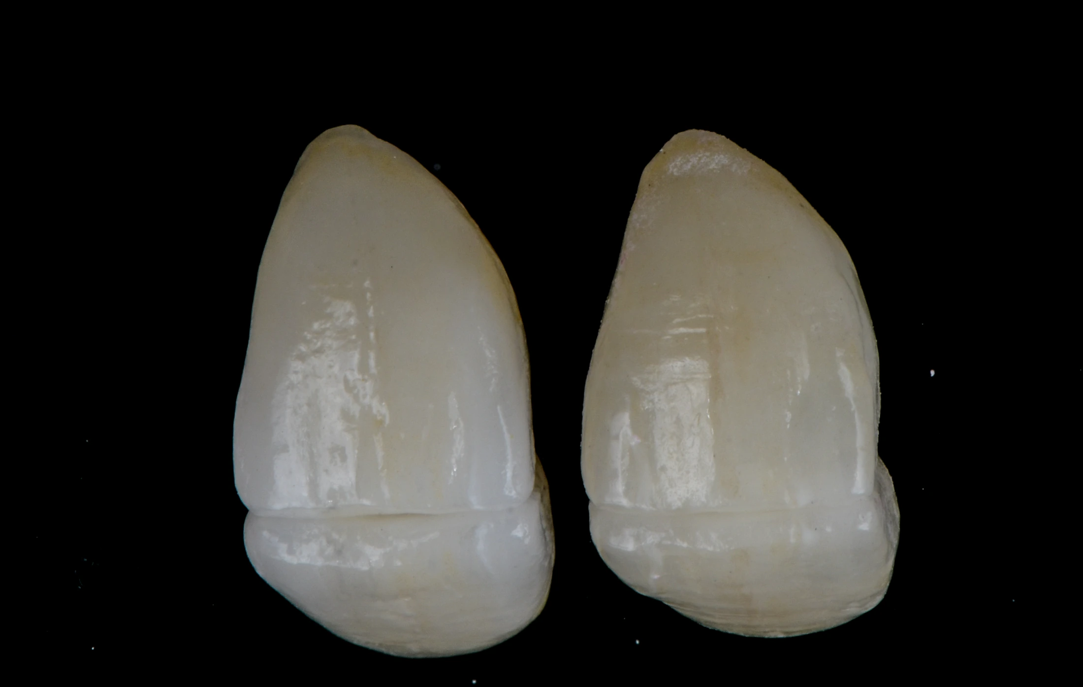
Intermediate lateral-incisor form with A1 and A2 shade-reference tabs.
03

Later clinical views

After: matching opposite-side retracted lateral view with teeth separated

Back to all cases

LET’S WORK TOGETHER

A considered result starts
with a conversation.

Discuss your next case

FROM THE LABORATORY

Follow the craft.

Explore Natalie’s Instagram grid for ceramic details, recent work and life in the laboratory.

Pink diagnostic wax-up from the laboratory collection Side-by-side restoration forms from the laboratory collection Natural smile from the clinical case collection

Selected photography from the website collection. The links open Natalie’s Instagram grid.